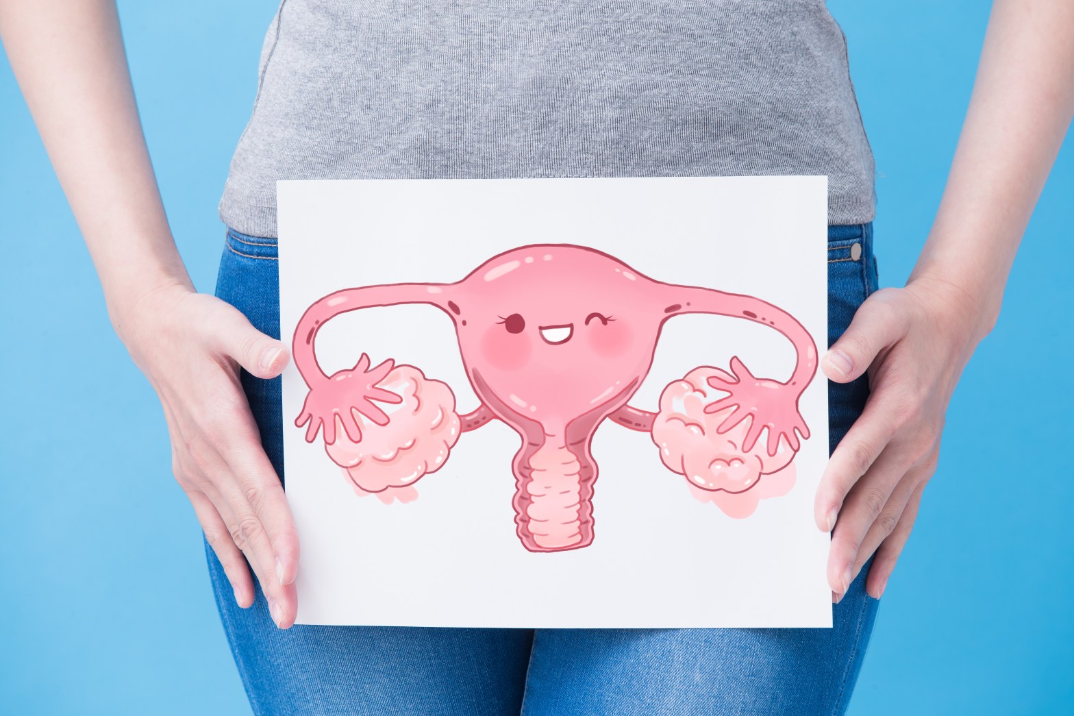
(Featured Image: https://stock.adobe.com/uk/images/woman-take-uterus-billboard/195321828?prev_url=detail&asset_id=192401061 (Licensed))
Dear Reader,
In my last post, I asked what color ribbon did you think represented endometriosis –
It’s Yellow.

Now, before we dive into the subject, I just want to cover the anatomy of the uterus and the endometrium for a better understanding of this condition.

The female reproductive system is divided into upper and lower genital tracts. The upper genital tract consists of the uterus, ovaries, and Fallopian tubes. The lower genital tract, in turn, consists of the cervix, vagina, and the external genitals, which includes the labia and the clitoris.[1,2,6]

[1,4,6,8]
The uterus consists of three layers:
- The thin outer layer called the Perimetrium or the serosa- thin layer of tissue surrounding the uterus.
- The middle smooth muscle layer, called the Myometrium– makes up most of the uterine volume and consists of smooth muscle cells.
- The innermost layer called the Endometrium– the most active layer that responds to cyclic ovarian hormone changes, essential for both menstrual and reproductive functions. It becomes thicker before ovulation and disintegrates before menstruation. [5,8]
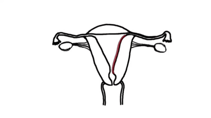
That is, in the absence of a pregnancy, the upper functional layer of the endometrium breaks down. It is shed into the uterine cavity during menstruation before flowing out the vagina.[12]
The Structure of Endometrium can be divided into two layers:[3,12]
- The layer nearest to the open cavity of the uterus is called the Functionalis, which is further broken down into compact and spongy zones. The functionalis zone undergoes significant dramatic changes throughout the menstrual cycle, and it is this zone that sheds itself every 28 days in a non-pregnant uterus.
- The second layer of the endometrium is the Basal zone. This zone contains the supporting vasculature, i.e. the Uterine artery. This zone undergoes much less damage during the menstrual cycle. It is thought to be primarily responsible for the regeneration of the endometrium as early as the second day of the menstrual bleeding.
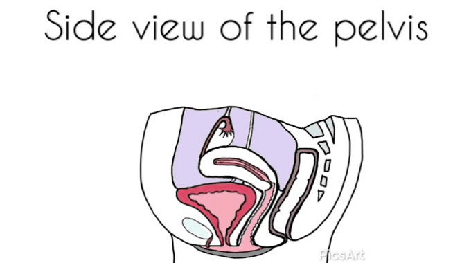
Let’s look at the side on view of the pelvis:
[1,2,6]
You can see the Uterus and the vagina, and then to the top, you can see one of the ovaries and the fallopian tube (Uterine tube), and you can see how they relate to other structures like the bladder and the rectum.[11,1,6]
The little space between the uterus and the rectum is called the Recto-uterine Pouch or Pouch of Douglas.[1]
The endometrial sites can commonly settle near or outside the uterus like the ovaries, uterine ligaments (mostly broad and uterosacral ligaments), the pouch of Douglas, and fallopian tubes.
The lesions have also been found outside the pelvic region, including the gastrointestinal tract, lungs, diaphragm, abdomen, and pericardium (Layer covering the heart).[3,7]
Did you know that it took 23 years for the famed celebrity model Padma Lakshmi to be taken seriously before she was finally diagnosed with endometriosis! [13]
Until next time,
With love,
Palasha.
References:
- Primal Pictures Ltd. (2001). Anatomy.tv. London: Primal Pictures.
- Drake, R., Vogl, W., Mitchell, A., Tibbitts, R., Richardson, P., & Gray, H. Gray’s anatomy for students (3rd ed., p. 177).
- Olive, D. (2005). Endometriosis in clinical practice. London: Taylor & Francis.
- Ellis, H. (2011). Anatomy of the uterus. Anaesthesia & Intensive Care Medicine, 12(3), 99-101.
- The uterus. (2019). Retrieved 11 November 2019, from https://anatomy-medicine.com/endocrine-system/101-the-uterus.html
- Chaurasia, B., Garg, K., Mittal, P., & Chandrupatla, M. (2017). BD Chaurasia’s human anatomy (Volume II). New Delhi: CBS Publishers & Distributors Pvt Ltd.
- Vercellini P, Viganò P, Somigliana E, Fedele L: Endometriosis: pathogenesis and treatment. Nat Rev Endocrinol. 2013, 10:261-275. 10.1038/nrendo.2013.255
- Gossman W, Fagan SE, Sosa-Stanley JN, et al. Anatomy, Abdomen and Pelvis, Uterus. [Updated 2019 Jul 11]. In: StatPearls [Internet]. Treasure Island (FL): StatPearls Publishing; 2019 Jan-. Available from: https://www.ncbi.nlm.nih.gov/books/NBK470297/
- Beth Smith’s answer to Have you ever had a medical condition or disease that doctors misdiagnosed? – Quora. (2019). Retrieved 28 December 2019, from https://qr.ae/TSU82z
- Olive DL, Pritts EA. Treatment of endometriosis. N Engl J Med.2001;345(4):266–275.oi:10.1056/NEJM200107263450407
- Ellis, H. (2011). Anatomy of the uterus. Anaesthesia & Intensive Care Medicine, 12(3), 99-101.
- Diamond, Michael P., and Kevin G. Osteen. Endometrium and Endometriosis. Oxford: Blackwell Science, 1997. Print.
- Practical Pain Management. (2020). Padma Lakshmi Talks to PPM about Living with the Emotional and Physical Toll of Endometriosis. [online] Available at: https://www.practicalpainmanagement.com/patient/conditions/pelvic-pain/story-padma-lakshmi-talks-ppm-about-living-emotional-physical-toll [Accessed 1 Jan. 2020].
Featured Image: https://stock.adobe.com/uk/images/woman-take-uterus-billboard/195321828?prev_url=detail&asset_id=192401061 (Licensed)

Very informative
LikeLike
Its informative. Like. I heard about endometrium.
LikeLike