⬻Ease from Disease II-B⤖

(Featured Image: https://stock.adobe.com/images/pelvic-floor/313483255 (Licensed))


3ln37o
[Courtesy of Imgflip.com]

Dear Reader,

Welcome to the final blog post of this Endometriosis journey, where I want to answer the question, What constitutes in the pelvic rehabilitation session for Endometriosis patient?

Before saying anything further, I would like to state that Physiotherapy management is individualized.

Since Chronic pelvic pain in women suffering from this sinister pathology may be a manifestation of central sensitization, regardless of endometriosis-specific factors[4], Pain Management is one of the first lines of intervention.

Addressing the complexity of pain experiences physio-therapeutically means assessing and managing local tissue issues within a broader context of sensitised protective mechanisms resulting from central nervous system (CNS) sensitivity.[7,8] 

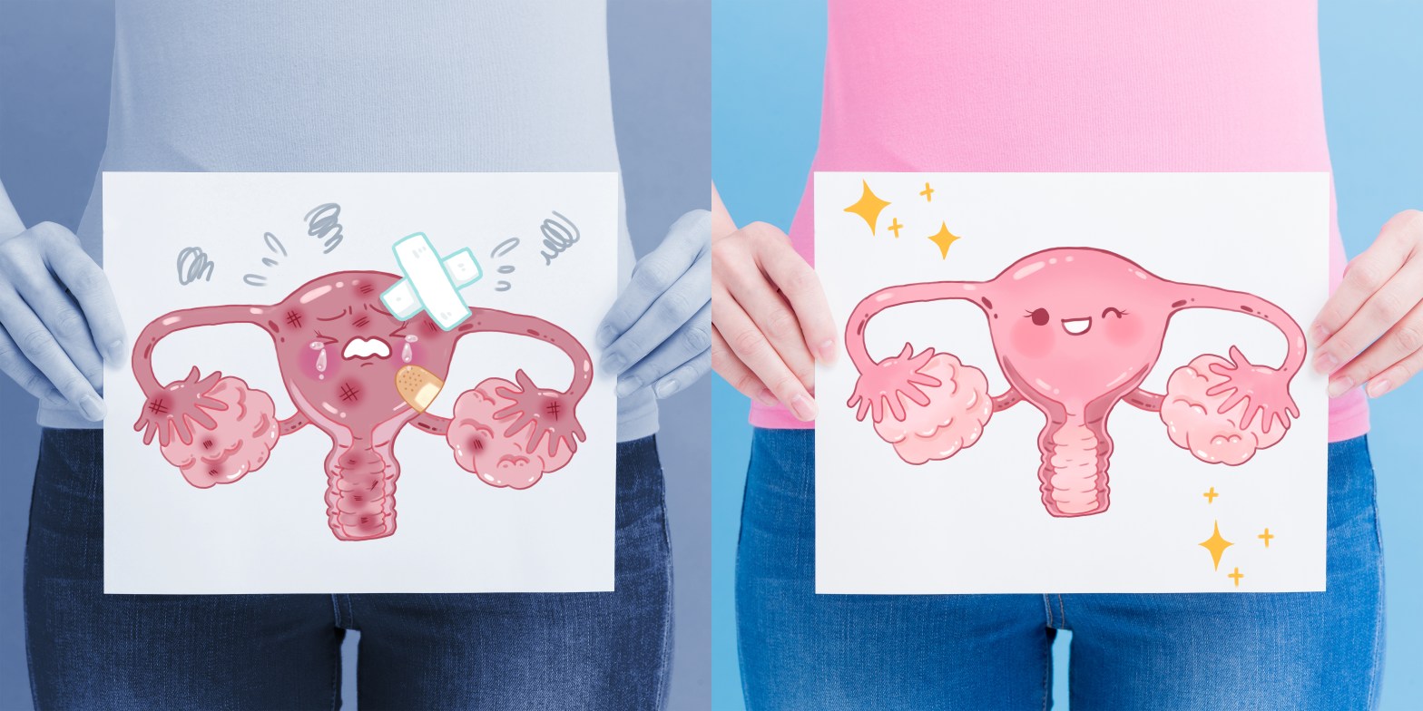
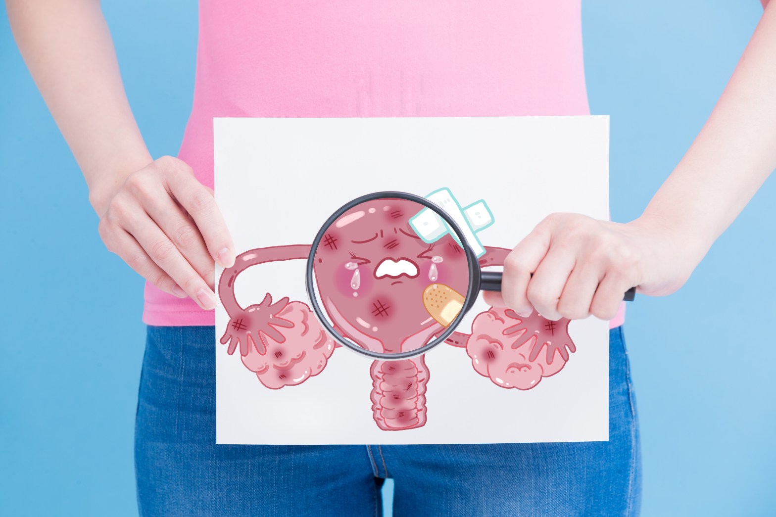
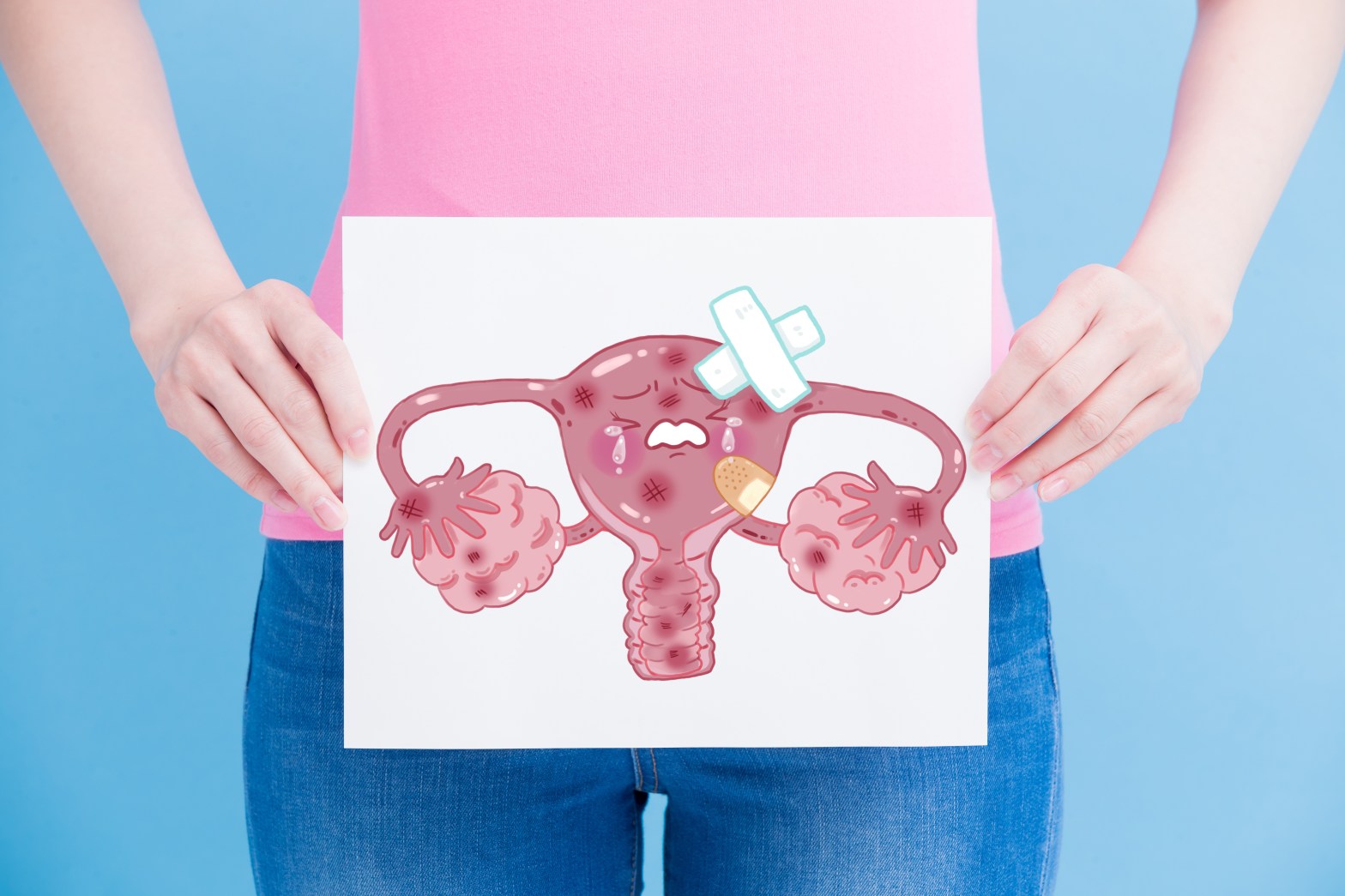
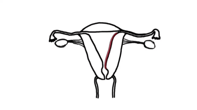
Many women have never seen images of genitalia or looked at their own vulva.[12]

Demystifying a woman’s genitalia is powerful and potentially achieved via education about anatomy, function, and vulval care. The need to normalize varied presentations as media images are very homogenized. Mirrors, pictures, and visualization may be helpful for some.[12]

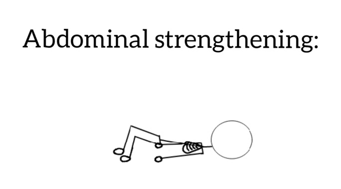
Now the pelvic physiotherapy rehabilitation usually consists of posture, core stabilization (like Pilates exercises), stretching, strengthening, flexibility, coordination exercises. [15]

20200112_171220

Khadiga et al. (2019) found out that Pilates was effective for treating pain and lowering blood cortisol levels in women with pelvic pain. [26]  Exercises like Pilates curl, roll-up, rolling like a ball, single straight leg stretch, double leg stretch, teaser 1, Pilates pushups, shoulder bridge, single leg kick were used.

20200113_105809
[26]

Manual therapy has the potential to reduce medication requirements in women with Chronic Pelvic Pain;[9] however, it may need delaying until some down-regulation of a sensitized central nervous system has occurred.[8]

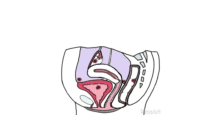
The rationale for the manual physical therapy protocol: To create micro failure of the attachments of collagen cross-links [16] since previous medical studies [17,18,19] have shown that endometriosis is frequently accompanied by adhesions.

Individualization of treatment occurred as the therapist focused on the adhesion areas, engaging the shortened soft tissues until the tissues often, indicating the release of cross-links.[19] Some of the Examples of the (Wurn’s Technique) Pelvic Manual Therapy are as follows:

20200112_223439 (1)
[19,20]

As shown in the first manual therapy example, the therapist engages the uterine fundus and sidewalls and tractions them to the left. To assist and improve the mobility of the soft tissues, the therapist may release the tension of the traction either suddenly or gradually, depending on the desired effect.[20]

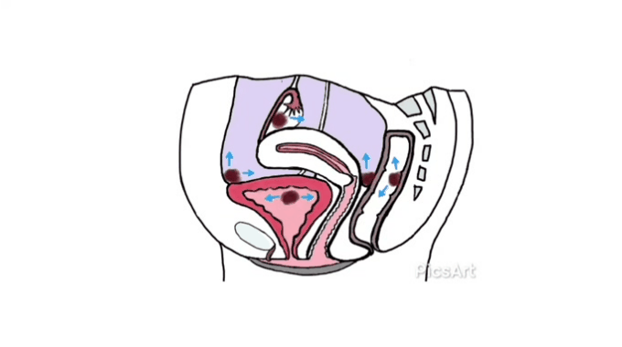
In the second example, the therapist uses the leg as a lever in one of the techniques to
release adhesions from the uterine fundus.

Bi et al. (2018) found that neuromuscular electrical stimulation (NMES) useful for the treatment of endometriosis-associated pain (EAP). [25] Commonly, pelvic health physios utilize digital techniques, with or without biofeedback and intravaginal devices- To improve:

  1. Awareness of contraction/relaxation
  2. proprioception
  3. Confidence
  4. Knowledge
  5. Self-awareness of the vagina and Pelvic Floor Muscles

In turn, reducing hypersensitivity and stretch soft tissues.[14]

Positive physical experiences can start to modify fear-based reactions.[12]

Gentle neural mobilization techniques can be beneficial: Adverse neural tension in pudendal, ilioinguinal, iliohypogastric, femoral, or obturator nerves may contribute to pelvic pain.[13]

Pulsed high-intensity laser therapy is an effective method of pain alleviation, reducing adhesions, and improving the quality of life in women with endometriosis. [27] Its sedative action may result from several mechanisms that involve its capacity to decrease the conduction of pain impulses and to raise the rate of production of substances in human tissue that mimic the action of morphine. [28]

In addition to being a physiotherapist, I am also a yoga instructor and an Aquatic Therapist. I feel it is essential to mention the alleviating effects of them in combination with Physiotherapy bring about psychological and physical benefits to the Endometriosis sufferers [15,21,22].

It is a recipe for healing!

Yoga is an effective, time-tested method for improving overall health and managing psychosomatic and chronic degenerative disorders.[23] Yoga therapy has shown a reduction in the severity and duration of pain in women with pelvic pain. Below is the sequence used by the Saxena and his team in the targeted group:

An external file that holds a picture, illustration, etc.Object name is IJY-10-9-g002.jpg
Saxena et al. (2017) used this above sequence of yoga regimes for pain relief.[21]

Water exercises/ Aquatic Therapy is also another effective method added to Pelvic Physical Therapy Rehabilitation.[15] Some of the examples of the water rehabilitation: Deep and shallow water walking and running, Aerobic exercises, flexibility exercises, power, coordination, speed, and agility in addition to other specific exercises for abdominal and pelvic muscles and thighs.[15,22]

In water with lower gravity power and hydrostatic resistance as well as the lower load on joints and muscle, muscles, tendons, and ligaments can be easily strengthened. [22]

Schuch et al. (2014) found that moderate intensity, water-based exercise improved physical and psychological domains of Quality of Life, depressive symptoms, aerobic capacity, and muscular strength of women.[24]

Posture and movement should be dynamic and varied, rather than static and bracing. Exercise has benefits beyond physical fitness, including regulating sleep, improving energy, mood, and inflammation.[14]

A quick summary of the physical physiotherapy interventions:

20200112_103036

I would like to conclude that, during my study of this complex world of Endometriosis, I found that there are very few articles dedicated to it.

That being said, I am grateful for these wonderful researchers researching in this field and I hope to join their team one day.

I owe this to the 176 million women out there.

I want to thank you, my dear Reader!

You have taken one step further in bringing out Awareness of this horrible condition. Thank you for taking the time, and I hope you enjoyed reading my blog as much as I loved writing it.

Please do share, comment, and like.

Let’s together fight in this stigma of pelvic conditions.

One awareness at a time.

Together.

With love,

Palasha.

References:

  1. Larbi, M., and Larbi, M. (2019). My endometriosis was so agonizing I burned myself to distract from the pain. [online] The Sun. Available at: https://www.thesun.co.uk/fabulous/9047918/endometriosis-agony-physiotherapy/ [Accessed 5 Jan. 2020].
  2. HE Bloomfield, A Olson, N Greer, et al. Screening pelvic examinations in asymptomatic, average-risk adult women: an evidence report for a clinical practice guideline from the American College of Physicians. Ann Intern Med. 2014;161(1):46-53
  3. Buggio, L., Barbara, G., Facchin, F., Frattaruolo, M. P., Aimi, G., & Berlanda, N. (2017). Self-management and psychological-sexological interventions in patients with endometriosis: strategies, outcomes, and integration into clinical care. International Journal of Women’s Health, Volume 9, 281–293. DOI:10.2147/ijwh.s119724
  4. P Stratton, I Khachikyan, N Sinai, et al. Association of chronic pelvic pain and endometriosis with signs of sensitization and myofascial pain. Obstet Gynecol. 2015;125(3):719-28.
  5. Menstruation pulls the trigger for inflammation and pain in endometriosis. Laux-Biehlmann A, D’Hooghe T, Zollner TM. Trends Pharmacol Sci. 2015;36:270–276
  6. Alimi, Y., Iwanaga, J., Loukas, M., & Tubbs, R. S. (2018). The Clinical Anatomy of Endometriosis: A Review. Cureus10(9), e3361. DOI:10.7759/cureus.3361
  7. Central Sensitization, I: Pain Physiology and Evaluation for the Physical Therapist. Journal of Women’s Health Physical Therapy. 2011;35(3):103-13.
  8. J Nijs, B Van Houdenhove, RA Oostendorp. Recognition of central sensitization in patients with musculoskeletal pain: Application of pain neurophysiology in manual therapy practice. Man Ther. 2010;15(2):135-41.
  9. Anderson, RH Harvey, D Wise, et al. Chronic pelvic pain syndrome: reduction of medication use after pelvic floor physical therapy with an internal myofascial trigger point wand. Appl Psychophysiol Biofeedback. 2015;40(1):45-52.
  10. C Allaire, T Aksoy, M Bedaiwy, et al. An interdisciplinary approach to endometriosis-associated persistent pelvic pain. Journal of Endometriosis and Pelvic Pain Disorders. 2017;9(2):77-86.
  11. SJ Moore, SH Kennedy. K. The initial management of chronic pelvic pain. Green-top Guideline No. 41. Royal College of Obstetricians and Gynaecologists. 2012. Available from: www.rcog.org.uk/globalassets/documents/guidelines/gtg_41.pdf.
  12. S Hilton, C Vandyken. The Puzzle of Pelvic Pain—A Rehabilitation Framework for Balancing Tissue Dysfunction and Central Sensitization, I: Pain Physiology and Evaluation for the Physical Therapist. Journal of Women’s Health Physical Therapy. 2011;35(3):103-13
  13. C Vandyken, S Hilton. The Puzzle of Pelvic Pain. Journal of Women’s Health Physical Therapy. 2012;36(1):44-54.
  14. Kirkaldy, E. (2020). Physio? But I’ve got endometriosis! – O&G Magazine. [online] O&G Magazine. Available at: https://www.ogmagazine.org.au/21/2-21/physio-but-ive-got-endometriosis/#easy-footnote-bottom-56-9057 [Accessed 12 Jan. 2020].
  15. Vural, M. (2018). Pelvic pain rehabilitation. Turkish Journal of physical medicine and rehabilitation64(4), 291–299. DOI:10.5606/tftrd.2018.3616
  16. Threlkeld AJ. The effects of manual therapy on connective tissue. Phys Ther 1992; 72: 893-902
  17. Parker JD, Sinai N, Segars JH, et al. Adhesion formation after laparoscopic excision of endometriosis and lysis of adhesions. Fertil Steril 2005; 84: 1457-61.
  18. Luciano DE, Roy G, Luciano AA. Adhesion reformation after laparoscopic adhesiolysis: where, what type, and in whom they are most likely to recur. J Minim Invasive Gynecol 2008; 15: 44-8.
  19. Wurn, B. F., Wurn, L. J., Patterson, K., King, C. R., & Scharf, E. S. (2011). Decreasing Dyspareunia and Dysmenorrhea in Women with Endometriosis via a Manual Physical Therapy: Results from Two Independent Studies. Journal of Endometriosis, 3(4), 188–196. https://doi.org/10.5301/JE.2012.9088
  20. Wurn, L. J., Wurn, B. F., King, C. R., Roscow, A. S., Scharf, E. S., & Shuster, J. J. (2004). Increasing orgasm and decreasing dyspareunia by a manual physical therapy technique. MedGenMed: Medscape general medicine6(4), 47.
  21. Saxena, R., Gupta, M., Shankar, N., Jain, S., & Saxena, A. (2017). Effects of yogic intervention on pain scores and quality of life in females with chronic pelvic pain. International journal of yoga10(1), 9–15. DOI:10.4103/0973-6131.186155
  22. Rezvani, S., Taghian, F., & Valiani, M. (2013). The effect of aquatic exercises on primary dysmenorrhoea in nonathlete girls. Iranian journal of nursing and midwifery research18(5), 378–383.
  23. Trakroo, M., Bhavanani, A. B., Pal, G. K., Udupa, K., & Krishnamurthy, N. (2013). A comparative study of the effects of asana, pranayama, and asana-pranayama training on neurological and neuromuscular functions of Pondicherry police trainees. International journal of yoga6(2), 96–103. DOI:10.4103/0973-6131.113398
  24. Schuch, F. B., Pinto, S. S., Bagatini, N. C., Zaffari, P., Alberton, C. L., Cadore, E. L., … Kruel, L. F. M. (2014). Water-Based Exercise and Quality of Life in Women: The Role of Depressive Symptoms. Women & Health, 54(2), 161–175. DOI:10.1080/03630242.2013.870634
  25. Bi, X. L., & Xie, C. X. (2018). Effect of neuromuscular electrical stimulation for endometriosis-associated pain: A retrospective study. Medicine97(26), e11266. DOI:10.1097/MD.0000000000011266
  26. KHADIGA S. ABD EL-AZIZ, Ph.D., E., MOHAMAD F. ABO EL-ENEIN, M.D., A. (2019). Effect of Pilates Exercise on Primary Dysmenorrhea. The Medical Journal of Cairo University, 87(March), 1187-1192. DOI: 10.21608/mjcu.2019.53326
  27. Thabet, A. A. E.-M. & Alshehri, M. A. (2018). Effect of Pulsed High-Intensity Laser Therapy on Pain, Adhesions, and Quality of Life in Women Having Endometriosis: A Randomized Controlled Trial. Photomedicine and Laser Surgery, 36(7), 363–369. DOI:10.1089/pho.2017.4419
  28. Zati A, Valent A. Physical therapy: new technologies in rehabilitation medicine (translated to English). Edizioni Minerva Medica 2006:162–185

Featured Image: https://stock.adobe.com/images/pelvic-floor/313483255 (Licensed)


⬻Ease from Disease II-A⤖

Continued from the excepts of the last post:

Koga et al. proposed that the prolonged use of the medical therapy post-surgery (higher than 6 months) can prevent recurrence of painful cramps(dysmenorrhoea) in most patients, but has little to no effect in controlling recurrent chronic pelvic pain or painful intercourse (dyspareunia) [9].

Most people with Chronic Pelvic Pain or Endometriosis have adverse sexual effects, including severe deep dyspareunia, which is associated with pelvic floor muscle (PFM) muscle pain/myalgia regardless of stage/location of endometriosis. [10]

Just a quick interruption in the flow!

I would like to quickly mention the anatomy of the pelvic floor muscles since these are the ones getting affected in the pelvic floor myalgia condition.

A pelvic examination(as tolerated by the patient) by the pelvic physiotherapist can reveal which one of the pelvic floor muscles are spasmed or tight or weak. 

20200111_035838
[Courtesy of the app Anatomy for the Artist for the skeleton] [13]

Women have an increased prevalence of Pelvic Floor Myalgia and obturator internus spasm and increased muscle tightness(Hypertonicity).

Reduced flexibility, dyssynergia (uncoordinated and abrupt movements), altered motor control with a habitual holding pattern, and reduced capacity to relax their pelvic floor myalgia may also be present both in the voluntary and functional scenarios.[14,15]

Abnormal findings:

Apical breathing (upper chest breathing) with a braced abdominal wall is typical.

Positive Carnett’s sign may suggest an abdominal wall or nerve irritation components to pelvic pain.[3]

Positive Carnett’s sign: If the tenderness is unchanged or increased when abdominal muscles are tensed.

Pelvic floor myalgias may contract involuntarily in response to threat and occur without conscious awareness.[2]

FitzGerald and Kotarinos found the muscles of the pelvic floor are, in some cases, short, tender, and dysfunctional in women with chronic pelvic pain (CPP) [5].

Those dysfunctions may be due to the misalignment of pelvic structures. [6,7]. Those muscle imbalance patterns resulting from Chronic Pelvic Pain may create constant stretching of the pelvic floor muscles, leading to weakness and emergence of muscle spasms and/or trigger points that, then, exacerbate or prolong the pain.[4]

To conclude II-A:

20200112_021313
Musculoskeletal symptoms also include extra-pelvic and abdominal musculature.[1]
Chronic pelvic pain severity is associated with the abdominal wall and Pelvic floor muscle myalgia and observed in women with endometriosis.[4] [11,12]


Endometriosis can produce secondary musculoskeletal impairments that may be amenable to physical therapy intervention. [11,12]

Pelvic visceral disorders frequently produce lumbar, sacroiliac, and pelvic-floor referred pain, which can result in muscle spasms, trigger points, and connective tissue dysfunction. [11]

References:

  1. Doggweiler, R., Whitmore, K. E., Meijlink, J. M., Drake, M. J., Frawley, H., Nordling, J., … Tomoe, H. (2016). A standard for terminology in chronic pelvic pain syndromes: A report from the chronic pelvic pain working group of the international continence society. Neurourology and Urodynamics, 36(4), 984–1008. DOI:10.1002/nau.23072
  2. Van der Velde, J., & Everaerd, W. (2001). The relationship between involuntary pelvic floor muscle activity, muscle awareness, and experienced threat in women with and without vaginismus. Behaviour Research and Therapy, 39(4), 395–408. DOI:10.1016/s0005-7967(00)00007-3
  3. Allaire, C., Aksoy, T., Bedaiwy, M., Britnell, S., Noga, H. L., Yager, H., & Yong, P. J. (2017). An Interdisciplinary Approach to Endometriosis-associated Persistent Pelvic Pain. Journal of Endometriosis and Pelvic Pain Disorders, 9(2), 77–86. https://doi.org/10.5301/jeppd.5000284
  4. Jarrell, J. Curr (2011) Endometriosis and Abdominal Myofascial Pain in Adults and Adolescents. Pain Headache Rep 15: 368. https://doi.org/10.1007/s11916-011-0218-y
  5. Dos Bispo, A. P. S., Ploger, C., Loureiro, A. F., Sato, H., Kolpeman, A., Girão, M. J. B. C., & Schor, E. (2016). Assessment of pelvic floor muscles in women with deep endometriosis. Archives of Gynecology and Obstetrics, 294(3), 519–523. DOI:10.1007/s00404-016-4025-x
  6. Lukic A, Di Properzio M, De Carlo S, Nobili F, Schimberni M,
    Bianchi P, Prestigiacomo C, Moscarini M, Caserta D (2015)
    Quality of sex life in endometriosis patients with deep dyspareunia before and after laparoscopic treatment. Arch Gynecol
    Obstet. DOI:10.1007/s00404-015-3832-9
  7. Neville CE, Fitzgerald CM, Mallinson T, Badillo S, Hynes C, Tu F (2012) A preliminary report of musculoskeletal dysfunction in female chronic pelvic pain: a blinded study of examination findings. J Bodyw Mov Ther 16:50–56
  8. Tu FF, Holt J, Gonzales J, Fitzgerald CM (2008) Physical therapy
    evaluation of patients with chronic pelvic pain: a controlled
    study. Am J Obstet Gynecol 272(e1-272):e7
  9. Koga, K., Takamura, M., Fujii, T., & Osuga, Y. (2015). Prevention of the recurrence of symptoms and lesions after conservative surgery for endometriosis. Fertility and Sterility, 104(4), 793–801. DOI:10.1016/j.fertnstert.2015.08.026
  10. Orr, N. L., Noga, H., Williams, C., Allaire, C., Bedaiwy, M. A., Lisonkova, S., … Yong, P. J. (2018). Deep Dyspareunia in Endometriosis: Role of the Bladder and Pelvic Floor. The Journal of Sexual Medicine, 15(8), 1158–1166. DOI:10.1016/j.jsxm.2018.06.007
  11. Troyer, M. R. (2007). Differential Diagnosis of Endometriosis in a Young Adult Woman With Nonspecific Low Back Pain. Physical Therapy, 87(6), 801–810. DOI:10.2522/ptj.20060141
  12. Prendergast SA, Weiss JM. Screening for musculoskeletal causes of pelvic pain.
    Clin Obstet Gynecol. 2003;46:773–782.
  13. Primal Pictures Ltd. Anatomy.tv. London: Primal Pictures, 2001. Web.
  14. C Allaire, T Aksoy, M Bedaiwy, et al. An interdisciplinary approach to endometriosis-associated persistent pelvic pain. Journal of Endometriosis and Pelvic Pain Disorders. 2017;9(2):77-86.
  15. K Bo, HC Frawley, BT Haylen, et al. An International Urogynecological Association (IUGA)/International Continence Society (ICS) joint report on the terminology for the conservative and nonpharmacological management of female pelvic or dysfunction. Neurourol Urodynam. 2016;9999:1-24.


⬻Ease from the Disease I⤖

(Featured Image: https://stock.adobe.com/uk/images/woman-take-uterus-billboard/199738093?prev_url=detail (Licensed))

Dear Reader,

In the last post, I asked you to guess which month was world endometriosis awareness celebrated- It’s March.[1]

Now that endometriosis is confirmed, the treatment plan is focused on managing pain, trying to limit the progression of the implants, and addressing the associated issues with fertility.

20200110_073331
[12]
  • The European Society of Human Reproduction and Embryology guideline for Endometriosis recommends the use of non-Steroidal Anti-Inflammatory Drugs or other analgesics to reduce endometriosis-associated pain.[12]
  • The guideline recommends prescribing hormonal treatment—combined Oral Contraceptives, progestagens, anti-progestogens, or GnRH agonists—“as one of the options, as it reduces endometriosis-associated pain”[12]
20200110_080741.gif
[3,5,12]

If Non-Steroidal Anti-Inflammatory Drugs and hormonal contraceptives are ineffective, the next step is treatment with a
GnRH analog such as leuprolide or goserelin (Zoladex), aka ‘medical menopause.’[13]

However, the therapy causes adverse effects, such as hot flashes, night sweats, and
possible bone loss, in many women (basically inducing the side effects of menopause). [13]

Infertility is a major complication and cannot be treated via medical therapy, like all medical treatments available for endometriosis work by suppressing ovulation.[3,5]

Surgical management is the primary treatment as it can improve the patient’s probability of spontaneous conception or pave the path for in vitro fertilization (IVF) in patients with severe endometriosis [3,5].

Patients with severe pain refractory to medical therapy also can benefit from surgery, as shown by the pain relief experienced by up to 95% of patients who underwent laparoscopy to excise lesions [3,5].

Hysterectomy (a surgical procedure to remove the womb/uterus) has been suggested for women with severe, debilitating, and refractory endometriosis who do not wish to become pregnant and in whom other therapeutic measures have failed [3,6].

Sad Disclaimer Alert!

Endometriotic lesions that recur after surgery have been found to occur in the same vicinity as the previous lesions and patients who undergo conservative surgery have a higher likelihood of recurrence, as some small residual implants might remain post-operatively [4].

—end of the alert—

This is why many Endometriosis sufferers undergo numerous surgeries in their lifetime until menopause actually sets in.

In my next post, I have included a prologue of Ease from disease Part II to make sense of what I will convey in the last, final blog post.

I hope you can get the gist what I’m trying to convey. It can get a little bit overwhelming right now due to the terminology. If any questions are bubbling in mind, please do ask!

Until next time,

With love,

Palasha.

References:

  1. En.wikipedia.org. (2020). Worldwide Endometriosis March. [online] Available at: https://en.wikipedia.org/wiki/Worldwide_Endometriosis_March [Accessed 4 Jan. 2020].
  2. Alimi, Y., Iwanaga, J., Loukas, M., & Tubbs, R. S. (2018). The Clinical Anatomy of Endometriosis: A Review. Cureus10(9), e3361. DOI:10.7759/cureus.3361
  3. Hurt KJ. Philadelphia, PA: Wolters Kluwer Health; 2015. Pocket Obstetrics and Gynecology.
  4. Koga, K., Takamura, M., Fujii, T., & Osuga, Y. (2015). Prevention of the recurrence of symptoms and lesions after conservative surgery for endometriosis. Fertility and Sterility, 104(4), 793–801. DOI:10.1016/j.fertnstert.2015.08.026
  5. A focus on the medical management of endometriosis. Casper RF: Introduction. Fertil Steril. 2017;107:521–522.
  6. Kim, S., Falcone, T., & Flyckt, R. (2017). Surgical Management of Endometriosis in Patients with Chronic Pelvic Pain. Seminars in Reproductive Medicine, 35(01), 054–064. DOI:10.1055/s-0036-1597306
  7. Maheux-Lacroix, S., Nesbitt-Hawes, E., Deans, R., Won, H., Budden, A., Adamson, D., & Abbott, J. A. (2017). The endometriosis fertility index predicts live births following surgical resection of moderate and severe endometriosis. Human Reproduction, 32(11), 2243–2249. DOI:10.1093/humrep/dex291
  8. Prescott, J., Farland, L. V., Tobias, D. K., Gaskins, A. J., Spiegelman, D., Chavarro, J. E., … Missmer, S. A. (2016). A prospective cohort study of endometriosis and subsequent risk of infertility. Human Reproduction, 31(7), 1475–1482. DOI:10.1093/humrep/dew085
  9. Evans, S. F., Brooks, T. A., Esterman, A. J., Hull, M. L., & Rolan, P. E. (2018). The comorbidities of dysmenorrhea: a clinical survey comparing symptom profile in women with and without endometriosis. Journal of Pain Research, Volume 11, 3181–3194. DOI:10.2147/jpr.s179409
  10. Doggweiler, R., Whitmore, K. E., Meijlink, J. M., Drake, M. J., Frawley, H., Nordling, J., … Tomoe, H. (2016). A standard for terminology in chronic pelvic pain syndromes: A report from the chronic pelvic pain working group of the international continence society. Neurourology and Urodynamics, 36(4), 984–1008. DOI:10.1002/nau.23072
  11. Orr, N. L., Noga, H., Williams, C., Allaire, C., Bedaiwy, M. A., Lisonkova, S., … Yong, P. J. (2018). Deep Dyspareunia in Endometriosis: Role of the Bladder and Pelvic Floor. The Journal of Sexual Medicine, 15(8), 1158–1166. DOI:10.1016/j.jsxm.2018.06.007
  12. Eshre.eu. (2013). Endometriosis guideline. [online] Available at: https://www.eshre.eu/Guidelines-and-Legal/Guidelines/Endometriosis-guideline.aspx [Accessed 6 Jan. 2020].
  13. Schrager, S., Falleroni, J., & Edgoose, J. (2013). Evaluation and treatment of endometriosis. Am Fam Physician87(2), 107-113.

⬻Diagnosing Endometriosis⤖

(Featured Image: https://stock.adobe.com/uk/images/woman-take-uterus-billboard/196513325?prev_url=detail (Licensed))

“My surgery was over in about 5 hours. Weeks later, I felt like a new person. He shared with me a video of my surgery, and for the first time, I could see the twisted mess inside me that endometriosis had made. And then I watched him remove it all. It was cathartic.”[1]

[Courtesy of Imgflip.com]

Dear Reader,

Did you know that the sufferers of Endometriosis are called as Endo-warriors? American pop singer Halsey began this movement using the term #Endowarriors to raise awareness on the social media platform of Instagram.[3]

In today’s post, I want to talk about the different ways Endometriosis can be diagnosed.

Endometriosis is typically defined by

Its histology:

Extra-uterine lesions consisting of endometrial glands, endometrial stroma, and/or hemosiderin-laden macrophages. [7]

Based on location and depth:

Lesions are further described as superficial peritoneal lesions, ovarian endometrioma, or deep endometriosis.[2,7]

Among those who ultimately receive a successful definitive diagnosis, contemporary literature describes delays from symptom onset to diagnosis ranging from 4 to 11 years.[10, 11]

Moreover, although the evidence is limited, failure of timely diagnosis and adequate management may foster disease progression and adhesion formation that may compromise fertility and increase the risk of central sensitization and chronic pelvic pain.[9,13]

Diagnosis

Anamnesis (a patient’s account of their medical history)

20200109_131001

Listening to the patient. Carry on a detailed anamnesis languidly. This simple action gives us the best approach to the disease. She has so much to tell, to show with her face and expression. In most cases, the disease can be understood just by listening.[2]

The omnipresent symptom is pain:

20200107_170359
[2,4,5]

There are many other pain presentations that nobody even thinks of until confronted with an endometriosis patient who, incidentally, has exactly “that type of pain.” Eguardo Rolla (2019) put forth a case of a young girl that he and his team had operated last year, who had referred right shoulder pain at menstruation. At laparoscopy, a large diaphragmatic series of blue and red lesions were excised. She was relieved after surgery. [2,6]

A similar case was encountered by Singh et al. (2017). He used MRI for the clinical diagnosis of endometriosis successfully. [6]

Pelvic Examination

Data from comparative studies suggest that findings on physical examination can identify endometriosis with high accuracy.[14,15]

Bimanual pelvic palpation:

20200109_140954
[2,7,15,16]

Uterosacral ligament compromise or adhesions at the Douglas pouch➟Fixed uterine retroversion and Dyspareunia(painful sexual intercourse) [2,7]

Painful uterine mobilization is another typical sign of endometriosis.[2]

Rictus of pain cannot be avoided. It will tell you exactly where the pain is more intense, helping to clinically determine the extent of the disease. The careful and expert pelvic examination provides a lot of information at a little cost.[2]

A caveat to bimanual examination is that it may not be feasible for non−sexually active adolescents/young adults and may not identify early-stage, superficial disease.[7]

Imaging

Imaging can be a useful adjunct to clinical diagnostic measures, and transvaginal ultrasound improves accuracy when used adjunctively with symptoms, patient history, and/or physical findings.[2,7,18]

Ultrasound is particularly sensitive for detecting ovarian endometriomas and deep endometriosis. Transvaginal ultrasound approaches the sensitivity and specificity needed to replace surgery for endometrioma detection.[19]

Nonetheless, not all endometriosis will be visualized by imaging, and imaging cannot be used to rule out endometriosis.

Laparoscopy


Laparoscopy is the “gold standard” for the diagnosis of endometriosis. It certifies the presence of the disease and its extension. Utilizing tissue biopsies and their pathological analysis, the aggressiveness of the lesions can be determined. It is also the opportunity to perform the initial treatment of endometriosis.[2,7]

I would like to take an opportunity to quickly explain the stages of endometriosis:

20200109_154303.gif
[17,21]

Revised American Society for Reproductive Medicine classification of endometriosis{ASRM} classifies endometriosis from Stage I (minimal) to Stage IV (severe) based on the location and size of the lesions seen during the surgical procedure (usually during exploratory surgeries).[17]

Before I end this post, I would like you to guess which month World Endometriosis Awareness is celebrated?

Until next time,

With love,

Palasha.

References:

  1. Flutter Health. (2017). Kristy’s Story — Flutter Health. [online] Available at: https://www.flutterhealth.com/kristys-story [Accessed 2 Jan. 2020].
  2. Rolla, E. (2019). Endometriosis: advances and controversies in classification, pathogenesis, diagnosis, and treatment. F1000Research, 8, F1000 Faculty Rev-529. DOI:10.12688/f1000research.14817.1
  3. Reimel, E. (2020). Halsey Got Real About Her Battle With Endometriosis in a Powerful Message to Fellow #Endowarriors. [online] Glamour. Available at: https://www.glamour.com/story/halsey-got-real-about-her-battle-with-endometriosis-in-a-powerful-endowarrior-instagram [Accessed 3 Jan. 2020].
  4. Giudice L. C. (2010). Clinical practice. Endometriosis. The New England journal of medicine, 362(25), 2389–2398. DOI:10.1056/NEJMcp1000274
  5. Laux-Biehlmann, A., d’ Hooghe, T., & Zollner, T. M. (2015). Menstruation pulls the trigger for inflammation and pain in endometriosis. Trends in Pharmacological Sciences, 36(5), 270–276. DOI:10.1016/j.tips.2015.03.004
  6. Singh, A., Das, C. J., Das, B. K., & Gupta, A. K. (2017). The utility of diffusion-weighted imaging in diagnosing subdiaphragmatic endometriosis presenting as shoulder pain. The Indian journal of radiology & imaging, 27(3), 314–317. DOI:10.4103/ijri.IJRI_86_16
  7. Agarwal, S., Chapron, C., Giudice, L., Laufer, M., Leyland, N., Missmer, S., Singh, S., and Taylor, H. (2019). Clinical diagnosis of endometriosis: a call to action.
  8. N.P. Johnson, L. Hummelshoj, G.D. Adamson, et al. World Endometriosis Society consensus on the classification of endometriosis
    Hum Reprod, 32 (2017), pp. 315-324
  9. Unger, C. A., & Laufer, M. R. (2011). Progression of Endometriosis in Non-medically Managed Adolescents: A Case Series. Journal of Pediatric and Adolescent Gynecology, 24(2), e21–e23. DOI:10.1016/j.jpag.2010.08.002
  10. Moradi, M., Parker, M., Sneddon, A., Lopez, V., & Ellwood, D. (2014). Impact of endometriosis on women’s lives: a qualitative study. BMC Women’s Health, 14(1). DOI:10.1186/1472-6874-14-123
  11. Noah, K. E., Hummelshoj, L., Webster, P., d’ Hooghe, T., de Cicco Nardone, F., de Cicco Nardone, C., … Zondervan, K. T. (2011). Impact of endometriosis on quality of life and work productivity: a multicenter study across ten countries. Fertility and Sterility, 96(2), 366–373.e8. DOI:10.1016/j.fertnstert.2011.05.090
  12. As-Sanie, S., Harris, R. E., Harte, S. E., Tu, F. F., Neshewat, G., & Clauw, D. J. (2013). Increased Pressure Pain Sensitivity in Women With Chronic Pelvic Pain. Obstetrics & Gynecology, 122(5), 1047–1055. DOI:10.1097/aog.0b013e3182a7e1f5
  13. Coxon, L., Horne, A. W., & Vincent, K. (2018). Pathophysiology of endometriosis-associated pain: A review of pelvic and central nervous system mechanisms. Best Practice & Research Clinical Obstetrics & Gynaecology. DOI:10.1016/j.bpobgyn.2018.01.014
  14. Bazot, M., Lafont, C., Rouzier, R., Roseau, G., Thomassin-Naggara, I., & Daraï, E. (2009). Diagnostic accuracy of physical examination, transvaginal sonography, rectal endoscopic sonography, and magnetic resonance imaging to diagnose deep infiltrating endometriosis. Fertility and Sterility, 92(6), 1825–1833. DOI:10.1016/j.fertnstert.2008.09.005
  15. Hudelist, G., Ballard, K., English, J., Wright, J., Banerjee, S., Mastoroudes, H., … Keckstein, J. (2011). Transvaginal sonography vs. clinical examination in the preoperative diagnosis of deep infiltrating endometriosis. Ultrasound in Obstetrics & Gynecology, 37(4), 480–487. DOI:10.1002/uog.8935
  16. Hudelist, G., Oberwinkler, K. H., Singer, C. F., Tuttlies, F., Rauter, G., Ritter, O., & Keckstein, J. (2009). Combination of transvaginal sonography and clinical examination for preoperative diagnosis of pelvic endometriosis. Human Reproduction, 24(5), 1018–1024. DOI:10.1093/humrep/dep013
  17. American Society for Reproductive. (1997). Revised American Society for Reproductive Medicine classification of endometriosis: 1996. Fertility and Sterility, 67(5), 817–821. DOI:10.1016/s0015-0282(97)81391-x
  18. Nnoaham, K. E., Hummelshoj, L., Kennedy, S. H., Jenkinson, C., & Zondervan, K. T. (2012). Developing symptom-based predictive models of endometriosis as a clinical screening tool: results from a multicenter study. Fertility and Sterility, 98(3), 692–701.e5. DOI:10.1016/j.fertnstert.2012.04.022
  19. Bossuyt, P. M., Farquhar, C., Johnson, N., & Hull, M. L. (2016). Imaging modalities for the non-invasive diagnosis of endometriosis. Cochrane Database of Systematic Reviews. DOI:10.1002/14651858.cd009591.pub2
  20. Fassbender, A., Burney, R. O., F. O, D., D’Hooghe, T., & Giudice, L. (2015). Update on Biomarkers for the Detection of Endometriosis. BioMed Research International, 2015, 1–14. DOI:10.1155/2015/130854
  21. Alimi, Y., Iwanaga, J., Loukas, M., & Tubbs, R. S. (2018). The Clinical Anatomy of Endometriosis: A Review. Cureus10(9), e3361. DOI:10.7759/cureus.3361


⬻How does it occur?⤖

(Featured Image: https://stock.adobe.com/uk/images/woman-take-uterus-billboard/195321828?prev_url=detail&asset_id=192401061 (Licensed))

[Courtesy of Imgflip.com]

Dear Reader,

One question is burning at the back of the throat-

How does Endometriosis occur?

Well, due to the complexity of this condition, there is no certain pathogenesis to explain endometriosis.[2,3,6,7,14] Multiple hypotheses and theories have been proposed for the pathogenesis of endometriosis. However, each individual theory fails to account for all types of endometriotic lesions and is not mutually exclusive – therefore, multiple mechanisms are likely to be involved.[27] Some of the main theories that stand out today are-

  1. Retrograde menstruation theory
  2. Stem Cell Theory
  3. Lymphateous spread
  4. Genetic Theory
20200105_135008
The most popular theory of endometriosis is Sampson’s theory of retrograde menstruation. (meaning the backward flow of menstrual blood)[23,24]

Fun fact: The word endometriosis was introduced by Sampson in 1927.[1,20,23]

Though I always wondered since, in endometriosis, lesions are found outside of the uterus and elsewhere (organs). Shouldn’t endometriosis be named as Ecto-metriosis?

Research showed 76-90% of menstruating women experience retrograde menstrual flow.

In contrast, only approximately 10% suffer from endometriosis, there is more to this condition than retrograde menstrual flow, which is what led to the proposal of the stem cell theory. [6-8]

Stem cell theory 

Human endometrium regenerates on a cyclical basis each month, likely mediated by endometrial stem/progenitor cells. [17]

Stem cells are basic cells that can become almost any type of cell in the body

Progenitor cells are early descendants of stem cells

Women with endometriosis also exhibit an increase in the number of basalis fragments in their menstrual blood, which may account for disease pathogenesis when compared to healthy women who will also shed functionalis stem/progenitor cells. [17]

As endometrial progenitor cells are shed during the menstrual cycle, the retrograde menstruation theory was expanded, and it was established that these stem cells spread to the peritoneum via this process. [21]

Lymphovascular metastasis theory suggests that endometrial cells could spread to ectopic sites via the lymphatic and hematogenous spread, accounting for the presence of endometriosis in distant locations outside the pelvis.[6,33]

The genetic/epigenetic theory


Although there’s no particular endometriosis gene that’s been found [26]. Two-thirds of women with a diagnosis of the condition report having a family member suffering from endometriosis[13].

The risk of developing endometriosis is 6%–9% higher in first-degree relatives of women and 15% higher when they had severe disease [12-14]. In twin sisters, the prevalence and the age of onset of endometriosis are similar[15].

More recently, hereditary factors were estimated to account for 50% of endometriosis [16].

Now,

20200106_043208
Endometriotic lesions are governed by endocrine cycles and cyclic bleeding, just like endometrium. [24,28]

The ectopic endometrial tissue responds to hormonal stimulation and undergoes cyclic growth and shedding. Without a way to drain, this causes internal accumulation of blood. [2,26] Laux-Bielman et al. (2015) proposed the menstruating tissue as the trigger for inflammatory pain in endometriosis through the activation of innate immune cells and peripheral nerve endings.[28]

20200107_114433
[7,24,33]

The failure to remove fragments of menstrual effluent from the abdominal cavity induces excessive local inflammation and irritation.[7,24,33] 20200108_164853

Isolated cells from menstrual endometrial fragments as well as non-cellular medium prepared from menstrual effluent induce alterations in the epithelial layer of the membrane forming the lining of the abdominal cavity to damage it, creating its own adhesion sites around the peritoneum.[33]

20200108_175129
Fibronectin receptors may contribute to the adhesion of endometriotic cells during menstruation [7,24,33]

The contributions of endometriosis to infertility are likely multifactorial, including⬎

Impaired tubo-ovarian function: Adhesions around the ovaries and the fallopian tubes that either block the ovary from releasing the eggs or hold the tubes in abnormal positions that prevent the egg from moving along smoothly and reaching the uterus.

Ovarian endometrioma: can cause pelvic inflammation and damage the eggs or prevent effective ovulation. Thus reducing oocyte quality and endometrial receptivity to implantation.[24,32]

Whew!

🚬 Smog Alert🚬

Did you know that although smoking is deleterious to many other aspects of health, smoking is associated with a decreased risk of endometriosis in many research studies?[29,30,31] This is NOT in any way to encourage smoking as the harmful cons outweigh the to-dos, but it’s interesting to find this occurring, isn’t it?

Until next time,

With love,

Palasha

References:

  1. Sampson JA: Peritoneal endometriosis due to the menstrual dissemination of endometrial tissue into the peritoneal cavity. American Journal of Obstetrics & Gynecology. 1927;14(4):422–469. 10.1016/S0002-9378(15)30003-X
  2. Vercellini, P., Viganò, P., Somigliana, E. et al. Endometriosis: pathogenesis and treatment. Nat Rev Endocrinol 10, 261–275 (2014) DOI:10.1038/nrendo.2013.255
  3. Rolla, E. (2019). Endometriosis: advances and controversies in classification, pathogenesis, diagnosis, and treatment. F1000Research, 8, F1000 Faculty Rev-529. DOI:10.12688/f1000research.14817.1
  4. Orrin Hatch: This is nothing short of a public health emergency. (2019). Retrieved 26 December 2019, from https://www.cnn.com/2018/03/27/opinions/endometriosis-start-a-conversation-hatch-opinion/index.html
  5. My Life with Endometriosis – Samara’s Story – Wessex Fertility. (2019). Retrieved 25 December 2019, from https://www.wessexfertility.com/blog/my-life-with-endometriosis-samaras-story/
  6. Paul J.Q. van der Linden, Theories on the pathogenesis of endometriosis, Human Reproduction, Volume 11, Issue suppl_3, November 1996, Pages 53–65, https://doi.org/10.1093/humrep/11.suppl_3.53
  7. Carpinello OJ, Sondheimer LW, Alford CE, et al. Endometriosis. [Updated 2017 Oct 22]. In: Feingold KR, Anawalt B, Boyce A, et al., editors. Endotext [Internet]. South Dartmouth (MA): MDText.com, Inc.; 2000-. Available from: https://www.ncbi.nlm.nih.gov/books/NBK278996/
  8. Blumenkrantz JM, Gallagher N, Bashore RA, Tenckhoff, H. Retrograde menstruation in women undergoing chronic peritoneal dialysis. Obstet Gynecol 1981; 57:667-70.
  9. Yang, J., & Huang, F. (2014). Stem cell and endometriosis: new knowledge may be producing novel therapies. International journal of clinical and experimental medicine, 7(11), 3853–3858.
  10. Guerriero S, Mais V, Ajossa S, et al. : The role of endovaginal ultrasound in differentiating endometriomas from other ovarian cysts. Clin Exp Obstet Gynecol. 1995;22(1):20–2
  11. Guerriero S, Ajossa S, Minguez JA, et al. : Accuracy of transvaginal ultrasound for the diagnosis of deep endometriosis in uterosacral ligaments, rectovaginal septum, vagina, and bladder: Systematic review and meta-analysis. Ultrasound Obstet Gynecol. 2015;46(5):534–45. 10.1002/uog.15667
  12. Simpson, J.L., Elias, S., Malinak, L.R., and Buttram, V.C.J. Heritable aspects of endometriosis. I. Genetic studies. Am J Obstet Gynecol. 1980; 137: 327–331
  13. Coxhead, D. and Thomas, E.J. Familial inheritance of endometriosis in a British population. A case-control study. J Obstet Gynecol. 1993; 13: 42–44
  14. Kennedy, S. The genetics of endometriosis. J Reprod Med. 1998; 43: 263–268
  15. Moen, M.H. Endometriosis in monozygotic twins. Acta Obstet Gynecol Scand. 1994; 73: 59–62
  16. Sapkota, Y., Attia, J., Gordon, S.D., Henders, A.K., Holliday, E.G., Rahmioglu, N. et al. Genetic burden associated with varying degrees of disease severity in endometriosis. Mol Hum Reprod. 2015; 21: 594–602
  17. Cousins FL, O DF, Gargett CE. Endometrial stem/progenitor cells and their role in the pathogenesis of endometriosis. Best Pract Res Clin Obstet Gynaecol. 2018;50:27–38. DOI:10.1016/j.bpobgyn.2018.01.011
  18. The pathophysiology of endometriosis and adenomyosis: tissue injury and repair. Arch Gynecol Obstet, 280 (2009), pp. 529-538
  19. Deane JA, Gualano RC, Garrett CE. Regenerating endometrium from stem/progenitor cells: it is abnormal in endometriosis. Asherman’s syndrome and infertility? Curr Opin Obstet Gynecol. 2013;25:193-200.
  20. Benagiano, G., Brosens, I., & Lippi, D. (2014). The History of Endometriosis. Gynecologic and Obstetric Investigation, 78(1), 1–9. DOI:10.1159/000358919
  21. Endometrial stem/progenitor cells: the first 10 years. Gargett CE, Schwab KE, Deane JA. Hum Reprod Update. 2015;22:137–163
  22. Olive DL, Pritts EA. Treatment of endometriosis. N Engl J Med.2001;345(4):266–275.oi:10.1056/NEJM200107263450407
  23. Slender M, Srinivasan S. Pathogenesis of endometriosis: Look no further than John Sampson [published online ahead of print, 2019 Oct 24]. Reprod Biomed Online. 2019; S1472-6483(19)30783-7. DOI:10.1016/j.rbmo.2019.10.007
  24. Olive, D. (2005). Endometriosis in clinical practice. London: Taylor & Francis.
  25. Borghese, B., Zondervan, K.T., Abrao, M.S., Chapron, C., and Vaiman, D. Recent insights on the genetics and epigenetics of endometriosis. Clin Genet. 2017; 91: 254–264
  26. Alimi, Y., Iwanaga, J., Loukas, M., & Tubbs, R. S. (2018). The Clinical Anatomy of Endometriosis: A Review. Cureus, 10(9), e3361. DOI:10.7759/cureus.3361
  27. Dhesi, A. S., & Morelli, & S. S. (2015). Endometriosis: A Role for Stem Cells. Women’s Health, 35–49. https://doi.org/10.2217/WHE.14.57
  28. Laux-Biehlmann, A., d’ Hooghe, T., & Zollner, T. M. (2015). Menstruation pulls the trigger for inflammation and pain in endometriosis. Trends in Pharmacological Sciences, 36(5), 270–276. DOI:10.1016/j.tips.2015.03.004
  29. Sahin Ersoy G, Zhou Y, İnan H, Taner CE, Cosar E, Taylor HS. Cigarette Smoking Affects Uterine Receptivity Markers. Reprod Sci. 2017;24(7):989–995. DOI:10.1177/1933719117697129
  30. Simonelli A, Guadagni R, De Franciscis P, et al. Environmental and occupational exposure to bisphenol A and endometriosis: urinary and peritoneal fluid concentration levels. Int Arch Occup Environ Health. 2017;90(1):49–61. DOI:10.1007/s00420-016-1171-1
  31. Vaughan, S., Coward, J. I., Bast, R. C., Jr, Berchuck, A., Berek, J. S., Brenton, J. D., … Balkwill, F. R. (2011). Rethinking ovarian cancer: recommendations for improving outcomes. Nature reviews. Cancer, 11(10), 719–725. DOI:10.1038/nrc3144
  32. Carvalho, LF, Rossener, R, Azeem, A, Malvezzi, H, Abrao, Simoes M, Agrawal, A. From conception to birth – how endometriosis affects the development of each stage of reproductive life. Minerva Ginecol. 65(2), 181198 (2013).
  33. Laganà AS, Garzon S, Götte M, et al. The Pathogenesis of Endometriosis: Molecular and Cell Biology Insights. Int J Mol Sci. 2019;20(22):5615. Published 2019 Nov 10. DOI:10.3390/ijms20225615

Featured Image: https://stock.adobe.com/uk/images/woman-take-uterus-billboard/195321828?prev_url=detail&asset_id=192401061 (Licensed)

⬻Anatomy⤖

(Featured Image: https://stock.adobe.com/uk/images/woman-take-uterus-billboard/195321828?prev_url=detail&asset_id=192401061 (Licensed))

Dear Reader,

In my last post, I asked what color ribbon did you think represented endometriosis –

It’s Yellow.

Image result for yellow ribbon emoji

Now, before we dive into the subject, I just want to cover the anatomy of the uterus and the endometrium for a better understanding of this condition.

20191107_181318-1

The female reproductive system is divided into upper and lower genital tracts. The upper genital tract consists of the uterus, ovaries, and Fallopian tubes. The lower genital tract, in turn, consists of the cervix, vagina, and the external genitals, which includes the labia and the clitoris.[1,2,6]

project_1573843417949

[1,4,6,8]

The uterus consists of three layers:

  • The thin outer layer called the Perimetrium or the serosa- thin layer of tissue surrounding the uterus.
  • The middle smooth muscle layer, called the Myometrium– makes up most of the uterine volume and consists of smooth muscle cells.
  • The innermost layer called the Endometrium– the most active layer that responds to cyclic ovarian hormone changes, essential for both menstrual and reproductive functions. It becomes thicker before ovulation and disintegrates before menstruation. [5,8]

20200105_045119

That is, in the absence of a pregnancy, the upper functional layer of the endometrium breaks down. It is shed into the uterine cavity during menstruation before flowing out the vagina.[12]

The Structure of Endometrium can be divided into two layers:[3,12]

  • The layer nearest to the open cavity of the uterus is called the Functionalis, which is further broken down into compact and spongy zones. The functionalis zone undergoes significant dramatic changes throughout the menstrual cycle, and it is this zone that sheds itself every 28 days in a non-pregnant uterus.
  • The second layer of the endometrium is the Basal zone. This zone contains the supporting vasculature, i.e. the Uterine artery. This zone undergoes much less damage during the menstrual cycle. It is thought to be primarily responsible for the regeneration of the endometrium as early as the second day of the menstrual bleeding.

Let’s look at the side on view of the pelvis:

20200105_004822

[1,2,6]

You can see the Uterus and the vagina, and then to the top, you can see one of the ovaries and the fallopian tube (Uterine tube), and you can see how they relate to other structures like the bladder and the rectum.[11,1,6]

The little space between the uterus and the rectum is called the Recto-uterine Pouch or Pouch of Douglas.[1]

The endometrial sites can commonly settle near or outside the uterus like the ovaries, uterine ligaments (mostly broad and uterosacral ligaments), the pouch of Douglas, and fallopian tubes.

The lesions have also been found outside the pelvic region, including the gastrointestinal tract, lungs, diaphragm, abdomen, and pericardium (Layer covering the heart).[3,7]

Did you know that it took 23 years for the famed celebrity model Padma Lakshmi to be taken seriously before she was finally diagnosed with endometriosis! [13]

Until next time,

With love,

Palasha.

References:

  1. Primal Pictures Ltd. (2001). Anatomy.tv. London: Primal Pictures.
  2. Drake, R., Vogl, W., Mitchell, A., Tibbitts, R., Richardson, P., & Gray, H. Gray’s anatomy for students (3rd ed., p. 177).
  3. Olive, D. (2005). Endometriosis in clinical practice. London: Taylor & Francis.
  4. Ellis, H. (2011). Anatomy of the uterus. Anaesthesia & Intensive Care Medicine, 12(3), 99-101.
  5. The uterus. (2019). Retrieved 11 November 2019, from https://anatomy-medicine.com/endocrine-system/101-the-uterus.html
  6. Chaurasia, B., Garg, K., Mittal, P., & Chandrupatla, M. (2017). BD Chaurasia’s human anatomy (Volume II). New Delhi: CBS Publishers & Distributors Pvt Ltd.
  7. Vercellini P, Viganò P, Somigliana E, Fedele L: Endometriosis: pathogenesis and treatment. Nat Rev Endocrinol. 2013, 10:261-275. 10.1038/nrendo.2013.255
  8. Gossman W, Fagan SE, Sosa-Stanley JN, et al. Anatomy, Abdomen and Pelvis, Uterus. [Updated 2019 Jul 11]. In: StatPearls [Internet]. Treasure Island (FL): StatPearls Publishing; 2019 Jan-. Available from: https://www.ncbi.nlm.nih.gov/books/NBK470297/
  9. Beth Smith’s answer to Have you ever had a medical condition or disease that doctors misdiagnosed? – Quora. (2019). Retrieved 28 December 2019, from https://qr.ae/TSU82z
  10. Olive DL, Pritts EA. Treatment of endometriosis. N Engl J Med.2001;345(4):266–275.oi:10.1056/NEJM200107263450407
  11. Ellis, H. (2011). Anatomy of the uterus. Anaesthesia & Intensive Care Medicine, 12(3), 99-101.
  12. Diamond, Michael P., and Kevin G. Osteen. Endometrium and Endometriosis. Oxford: Blackwell Science, 1997. Print.
  13. Practical Pain Management. (2020). Padma Lakshmi Talks to PPM about Living with the Emotional and Physical Toll of Endometriosis. [online] Available at: https://www.practicalpainmanagement.com/patient/conditions/pelvic-pain/story-padma-lakshmi-talks-ppm-about-living-emotional-physical-toll [Accessed 1 Jan. 2020].

Featured Image: https://stock.adobe.com/uk/images/woman-take-uterus-billboard/195321828?prev_url=detail&asset_id=192401061 (Licensed)

⬻It’s all in your head⤖

(Featured Image: https://stock.adobe.com/uk/images/illustration-concept-of-pain-symptom-or-syndrome-in-pathologies-and-diseases-of-female-genitals-as-pms-endometriosis-anatomical-model-of-uterus-is-next-to-poster-on-which-written-in-red-word-pain/171284564 (Licensed))

“My Physician dismissed my debilitating period of pain for years. She would not prescribe a stronger painkiller as, ‘It works for most women,’ but it wasn’t working for me! I was throwing up for 3 days out of every 28, from this horrendous period pain equivalent to labor contractions.  ” [5]

[Courtesy by Imgflip.com]

Dear Reader,

Imagine this: You’re at a party. There are loads of people there. Disco lights going on- lights somehow missing you. You try talking to some people, but it seems that no one can hear you.

Suddenly, there’s a wound in your lower stomach (stab?) with blood spurting out. It’s an excruciating pain that’s continuous and doesn’t stop.

Yet no one sees you or hears you. The party keeps going on- disco lights all over the room, still missing you.

Two words- All Alone. That’s what your feeling.

project_1574514752176.gifThat’s how many, many women feel- alone. When they realize that bleeding buckets, excruciating pain following them isn’t healthy. They try to seek help and to diagnose this abnormality, only to be dismissed as normality or misdiagnosed.[6]

It’s not a funny business when a person tells you what you’re experiencing is normal when it’s not. It feels like the party situation- it’s like you are invisible.

What is the normality in this abnormality? It is the fact that it is conditioned in everyone’s brains that periods are supposed to be painful. But what if that ‘pain’ makes you want to cut your uterus out and kill yourself? [6-7]

Endometriosis.

A condition that {brace yourself} affects 1 in 10 women in reproductive years, that says approximately 176 million women in the world. Phew! [1-3]

What is Endometriosis, you say?

Good question.

Endometriosis is a condition where tissue similar to the lining of the uterus (the endometrial stroma and glands, which should only be located inside the uterus) is found elsewhere in the body. [1,4]

The very condition that is complex in nature to treat and to understand. Intellectuals and sufferers all over the world trying to understand what this condition is.


And if you see the side of the coin: A lack of awareness on the patient side can delay diagnosis tremendously.

Menstrual taboos still persist, whether by family or through sexual education, that period might be painful or uncomfortable. What they never understood was how painful (or not) an average period should be.

I’m Palasha, and I’m a physiotherapist. I come from a country where periods or anything related to women’s issues is a little stigmatized. [8]

I’m passionate about pelvic health, and I’m not afraid to talk about something which needs to be addressed and to normalize our pelvis. I mean, it is high time for that! Allow me to deliver the latest updated information on it and how, as a physiotherapist, I can play a role in treating it.

20200111_140946

Before we sign off, I would like to ask you a question. What color ribbon do you think represents Endometriosis?

Until then,

With lots of love,

Palasha.

References:

  1. org. (2019). Facts about endometriosis « Endometriosis.org. [online] Available at: http://endometriosis.org/resources/articles/facts-about-endometriosis/ [Accessed 19 Nov. 2019].
  2. Rogers PA, et al. Priorities for endometriosis research: recommendations from an international consensus workshop. Reprod Sci 2009;16(4):335-46.
  3. Adamson GD, et al. Creating solutions in endometriosis: global collaboration through the World Endometriosis Research Foundation. J of Endometriosis 2010;2(1):3-6.
  4. Kennedy, S., Bergqvist, A., Chapron, C., D’Hooghe, T., Dunselman, G., Greb, R., Hummelshoj, L., Prentice, A., and Saridogan, E. (2005). ESHRE guideline for the diagnosis and treatment of endometriosis. Human Reproduction, 20(10), pp.2698-2704.
  5. Jennette Wright’s answer to Has a doctor gave you a stupid diagnosis like ‘it’s in your head’ because he couldn’t admit he couldn’t find out? – Quora. (2019). Retrieved 28 December 2019, from https://qr.ae/TSOLqZ
  6. Thiagarajan, K (2019). ‘I’m trapped in the prison of my body’: The severe, chronic pain of endometriosis. [online] Scroll.in. Available at: https://scroll.in/pulse/872166/im-trapped-in-the-prison-of-my-body-the-severe-chronic-pain-of-endometriosis [Accessed 22 Nov. 2019].
  7. Ballard K, e. (2019). What’s the delay? A qualitative study of women’s experiences of reaching a diagnosis of endometriosis. – PubMed – NCBI. [online] Ncbi.nlm.nih.gov. Available at: https://www.ncbi.nlm.nih.gov/pubmed/?term=What%E2%80%99s+the+delay%3F+A+qualitative+study+of+women%E2%80%99s+experiences+of+reaching+a+diagnosis+of+endometriosis [Accessed 23 Nov. 2019].
  8. Mehedintu C, e. (2019). Endometriosis still a challenge. – PubMed – NCBI. [online] Ncbi.nlm.nih.gov. Available at: https://www.ncbi.nlm.nih.gov/pubmed/25408753 [Accessed 21 Nov. 2019].

Featured Image: https://stock.adobe.com/uk/images/illustration-concept-of-pain-symptom-or-syndrome-in-pathologies-and-diseases-of-female-genitals-as-pms-endometriosis-anatomical-model-of-uterus-is-next-to-poster-on-which-written-in-red-word-pain/171284564 (Licensed)

♥The Beginning♥

(Featured Image:  https://stock.adobe.com/uk/images/preface/261263903?prev_url=detail (Licensed))

Welcome!

Greetings to my blog,

A disclaimer needed to start,

   Intentions to make minds unclogged

It is a bit cheeky with moving art

It speaks nothing but the truth

All the gifs and artwork were done by yours truly (That’s me!)

This one’s dedicated to the women who lost their youth.

In Pain.

Table of Contents

  1. The Introduction
  2. Anatomy
  3. Theories of occurrence
  4. Diagnosis
  5. Treatment Part 1
  6. Treatment Part 2 A
  7. Treatment Part 2 B


Design a site like this with WordPress.com
Get started